
This condition is referred to as atrioventricular (AV) dissociation.
.jpg)
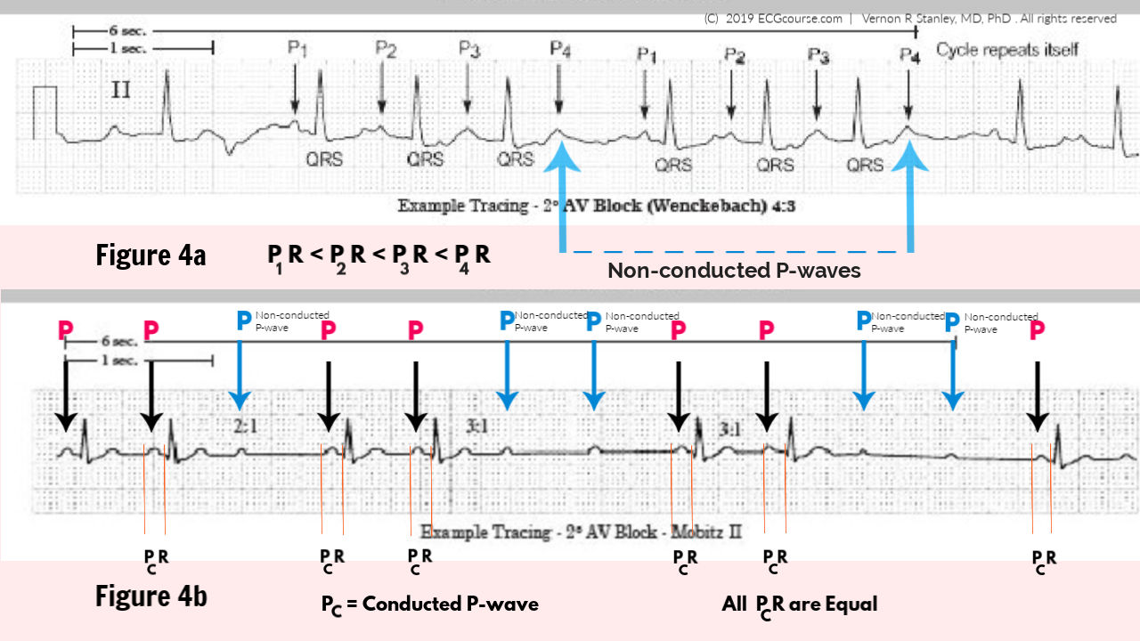
The atria and the ventricles are electrically dissociated from each other. In third-degree AV block no atrial impulses are conducted to the ventricles. Third-degree AV block (synonyms: complete heart block, AV dissociation, AV block III, AV block 3) Second-degree AV block (particularly Mobitz type 2) requires treatment. May also be referred to as Wenckebach block. In second-degree AV block some impulses are completely blocked, which means that not all P-waves are followed by QRS complexes. Second-degree AV-block occurs in the following two variants: Second-degree AV block (synonyms: AV block 2, AV block II, 2nd degree AV block) However, all impulses are conducted to the ventricles. First-degree AV block is rarely serious and may be left untreated in the vast majority of cases (exceptions are discussed later). By definition the PR interval is >0.22 s. The term block is somewhat misleading in this case, because first-degree AV block only implies that the conduction is abnormally slow. First-degree AV block (synonyms: AV block 1, AV block I, 1st degree AV block) First-, second- and third-degree AV block may all be diagnosed using the ECG. These conditions are referred to as atrioventricular (AV) blocks, which are subdivided according to the degree of block. Impulse conduction from the atria to the ventricles may be abnormally delayed or even blocked. Very strong parasympathetic input may lead to complete block of impulses. Sympathetic input causes increased impulse conduction ( bathmotropic effect), whereas parasympathetic input causes increased resistance in the atrioventricular node (additional slowing of the impulse). The atrioventricular (AV) node is richly innervated with sympathetic and parasympathetic fibers. Atrioventricular (AV) blocks occur due to dysfunction in the conduction system. Components of the ventricular conduction system and the temporal association between the ECG waveforms and impulse transmission through the heart. This is important since it optimizes the efficiency of the contraction. Refer to Figure 1. The rapid impulse transmission enables the majority of ventricular myocardium to be depolarized (more or less) simultaneously. Impulse conduction through the Purkinje system is very rapid due to the high abundance of gap junctions. From these bundles and fascicles, the Purkinje fibers sprout out into the myocardium. The left bundle branch is further divided into two fascicles. It causes a delay which gives the atria enough time to empty their blood into the ventricles, before ventricular contraction starts.Īfter leaving the atrioventricular node, the impulse continues through the His bundle which branches into the left and right bundle branch. Nevertheless, the slow impulse conduction through the atrioventricular node has a physiological purpose. Contractile cells and, in particular, Purkinje fibers, have an abundance of gap junctions which enables rapid impulse conduction. This is explained by the scarcity of gap junctions in the cells of the atrioventricular node. Impulse conduction through the atrioventricular node is slow. These structures conduct the atrial impulse to the ventricles. The AV system consists of the atrioventricular node (AV node) and the His-Purkinje system.

The atrioventricular (AV) conduction system and AV blocks Readers who are already familiar with AV blocks may skip to the subsequent articles discussing each type of AV block in detail: Below follows a general discussion on AV blocks, with emphasis on ECG characteristics and clinical features. There are three types of AV blocks, referred to as 1st degree AV block, 2nd degree AV block and 3rd degree AV block. In this article you will learn about the principles of atrioventricular (AV) blocks. Atrioventricular block (AV block): definition, causes, diagnosis & management


 0 kommentar(er)
